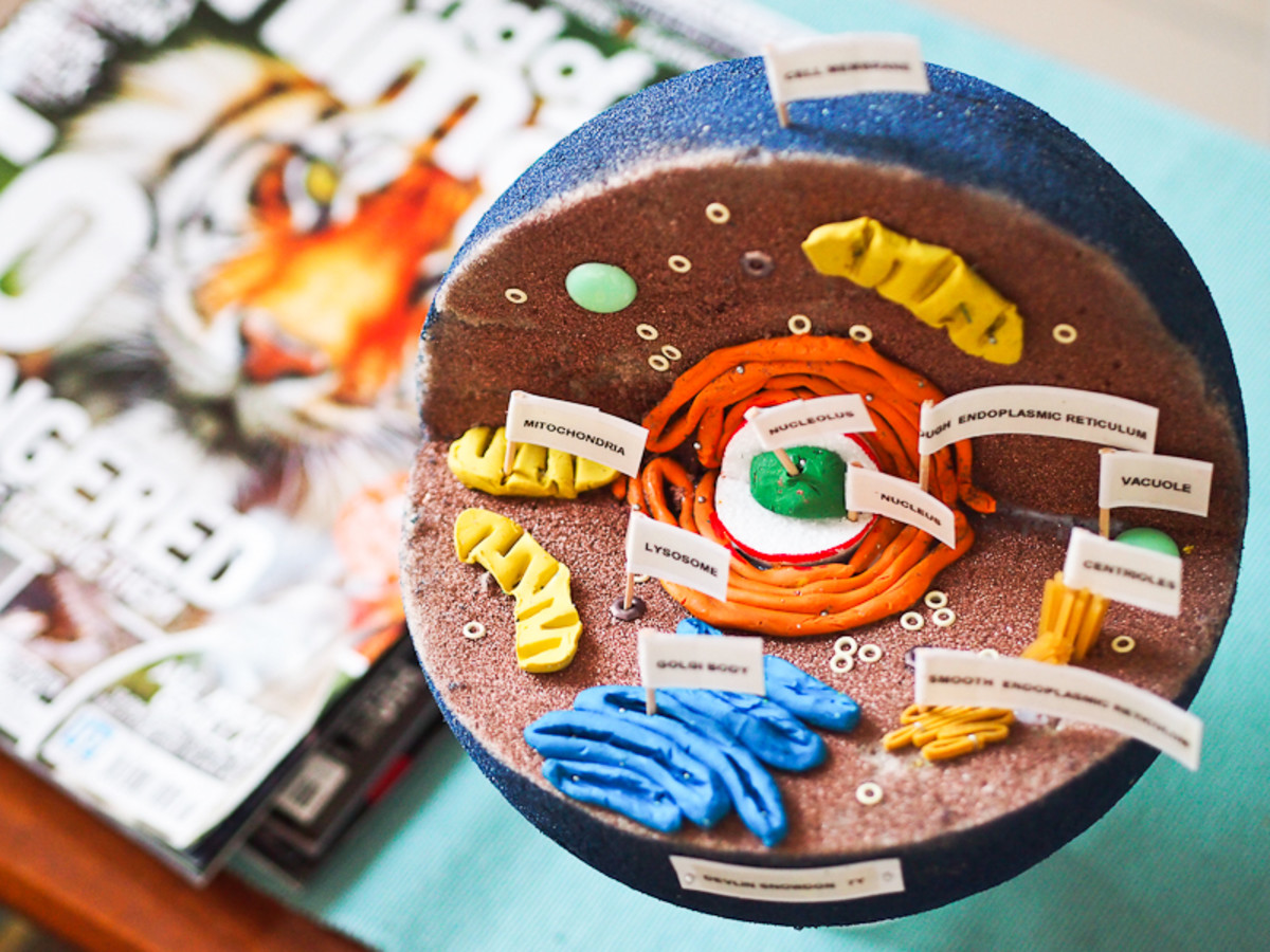
Labelled Diagram Of A White Blood Cell biology The cell from Latin cella meaning small room is the basic structural functional and biological unit of all known living organisms A cell is the smallest unit of life Cells are often called the building blocks of life The study of cells is called cell biology Cells consist of cytoplasm enclosed within a membrane which contains many biomolecules such as proteins and nucleic acids Labelled Diagram Of A White Blood Cell compound eye may consist of thousands of individual photoreceptor units or ommatidia ommatidium singular The image perceived is a combination of inputs from the numerous ommatidia individual eye units which are located on a convex surface thus pointing in slightly different directions
terrapsych ecology htmlThe glossary that follows assumes a definition of ecology the study of interactions between organisms and their environment much wider than what fits under the field s habitual statistical persona Ecofeminism and ecopsychology are mentioned for example as are terms from organic gardening and permaculture Labelled Diagram Of A White Blood Cell and Animal Cell Organelles The cells of eukaryotes protozoa plants and animals are highly structured These cells tend to be larger than the cells of bacteria and have developed specialized packaging and transport mechanisms that may be necessary to support their larger size cronodon BioTech Bacteria htmlBacteria singular bacterium are minute organisms that often consist of single cells like the rod shaped cell shown above which is about one thousandth of a millimetre mm in diameter that is one micrometre or about a tenth the diameter of
uq edu au School Science Lessons UNPh33 1 html3 84 2 Test a simple cell with different metals See Alligator Clips Commercial See Batteries Commercial See Voltmeters Commercial See diagram 33 3 1 Simple cell magnesium and copper Use alligator clips to connect a zinc strip to the negative terminal of a voltmeter and a copper strip to the positive terminal Labelled Diagram Of A White Blood Cell cronodon BioTech Bacteria htmlBacteria singular bacterium are minute organisms that often consist of single cells like the rod shaped cell shown above which is about one thousandth of a millimetre mm in diameter that is one micrometre or about a tenth the diameter of Service provided by the operator Business of LIFEX LIFEX records and stores knowledge such as medical big data its analysis results results obtained from it in block chains and provides useful information to stakeholders and commercial users
Labelled Diagram Of A White Blood Cell Gallery
Infected_RBC medium, image source: ocw.jhsph.edu

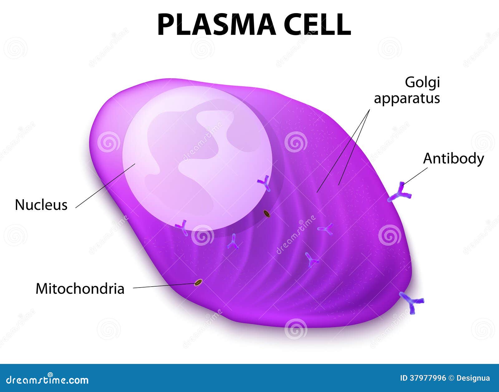
structure plasma cell b plasmocyte white blood cells secrete antibodies transported 37977996, image source: www.dreamstime.com
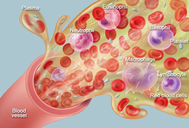
Blood cells 1031221, image source: www.motifolio.com

clip_image002 131, image source: www.biologydiscussion.com


labeled diagram of connective tissue df7a0db49b4bcd9811dd77bb1e3e3210, image source: anatomybody101.com
blood, image source: www.webmd.com
![]()
stock vector lymphocyte 394074688, image source: www.shutterstock.com

220px Platelet_structure, image source: en.wikipedia.org
mrs abrey lesson 14 blood 10 638, image source: www.slideshare.net

heme002, image source: pmgbiology.com
wpa0d1cb86_05_06, image source: www.sparklebox.co.uk

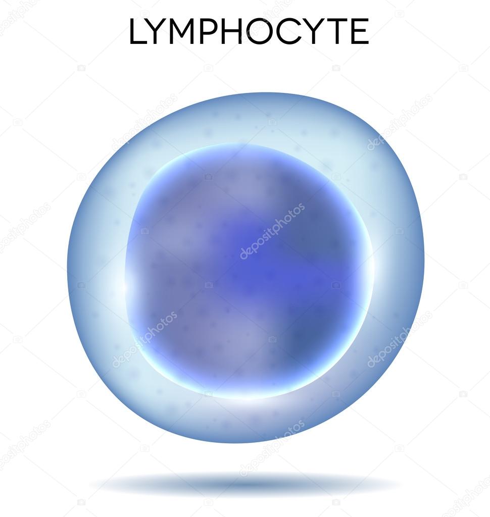
depositphotos_49757357 stock illustration human blood cell lymphocyte, image source: depositphotos.com

synapse labeled1 300x271, image source: thesalience.wordpress.com
lung lm1_med, image source: ib.bioninja.com.au

tonsil2, image source: legacy.owensboro.kctcs.edu

12636059_f520, image source: owlcation.com
clip_image0046, image source: www.biologydiscussion.com

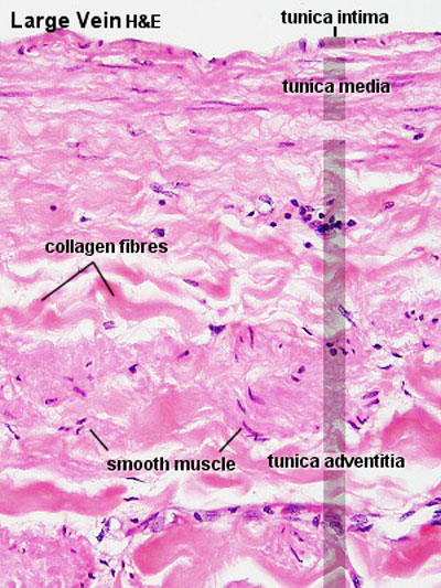
Vein_histology_02, image source: embryology.med.unsw.edu.au